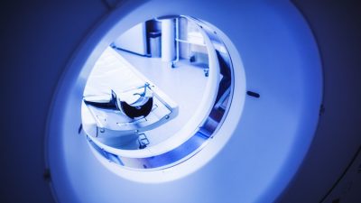
The first use of non-invasive fractional flow reserve-derived from CT coronary angiography (FFRct) in Australian patients is reported in our article, now published in Heart, Lung and Circulation [1]. This technology is currently TGA-approved in Australia, although not approved for use under Medicare.
Advances in CT coronary angiography currently offer the ability to assess both the anatomical and physiological aspects of coronary disease within the one scan. Non-invasive fractional flow reserve derived from CT coronary angiography (FFRct) is a technology first described in 2011 [2], pioneered by HeartFlow in the USA. Large multicentre global validation studies have since established its diagnostic performance and prognostic implications to be comparable with invasive fractional flow reserve. Across the US, Europe and Japan, and following regulatory approval, FFRct has evolved to become part ofmainstream clinical practice; in US (American Heart Association/American College of Cardiology) and UK (The National Institute for Health and Care Excellence [NICE] guidelines, FFRct is currently recommended for use in patients with stable recent onset chest pain.
How does FFRct work? Using the images of a routinely acquired CT coronary angiogram, a model of the coronary luminal tree is derived. Physiological assumptions are made regarding the viscosity of blood, as well as inlet and outlet flow and pressure, based on their observed relationship in accordance with vessel size and myocardial mass. These assumptions are then applied to the luminal model. Flow and pressure are derived using computational fluid dynamics across the entire coronary tree [3]. When compared with invasive fractional flow reserve, FFRct has high diagnostic performance. Importantly, it provides improved specificity for detection of vessel specific ischaemia compared with anatomical stenosis assessment using CT coronary angiography alone.
Our study included 109 patients who had undergone CT coronary angiography and invasive fractional flow reserve; the technology of FFRct was retrospectively applied. In this cohort of Australian patients, the diagnostic performance of FFRct was found to be comparable with the existing international literature, with demonstrated improvement in performance compared with CT coronary angiography alone for detection of vessel specific ischaemia.
[1]Chua A, Ihdayhid AR, Linde J, Sorgaard M, Cameron JD, Seneviratne S, Ko BS. Diagnostic Performance of CT-derived Fractional Flow Reserve in Australian Patients Referred for Invasive Coronary Angiography. Heart Lung Circ 2022; Article in press https://www.heartlungcirc.org/article/S1443-9506(22)00115-9/fulltext
[2]Koo BK, Erglis A, Doh JH, Daniels DV, Jegere S, Kim HS, et al. Diagnosis of ischemia-causing coronary stenoses by noninvasive fractional flow reserve computed from coronary computed tomographic angiograms. Results from the prospective multicenter DISCOVER-FLOW (Diagnosis of Ischemia-Causing Stenoses Obtained Via Noninvasive Fractional Flow Reserve) study. J Am Coll Cardiol. 2011;58:1989-97.
[3]Khav N, Ihdayhid AR, Ko B. CT-derived Fractional Flow Reserve (CT-FFR) in the Evaluation of Coronary Artery Disease. Heart Lung Circ 2020; 29: 1621-32.
Summary by co-author Brian Ko