A Clinical Case – what do you think?
by Andrew Martin
Mr A is a 72 year old who had a dual chamber permanent pacemaker implanted five years previously for symptomatic sinus node dysfunction – syncope with documented sinus pauses of 3-5 seconds duration. The right ventricular lead was inserted via the left cephalic vein and the right atrial lead via the left axillary vein with the generator sited within a pre- pectoral pocket. He is otherwise fit and well with no other co-morbidities, a structurally normal heart as assessed by echocardiography, and takes no regular medical therapies. His functional capacity is unlimited and he continues employment in a physical occupation.
He presents for routine assessment at the pacemaker clinic. His pacemaker is programmed DDDR, with rate range 50-140 per minute. With this programming his atrial pacing burden is 50% and his ventricular pacing burden is < 1%. As part of this interrogation, multiple episodes of non-physiological high frequency ‘noise’ sensed episodes have been recorded on the atrial channel. In addition, the atrial lead impedance is now > 3,000 Ohms, having previously been stable at 800-850 Ohms. Adduction of the left shoulder during the pacemaker interrogation was able to replicate the high frequency ‘noise’.
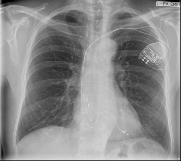
A chest x-ray has been obtained and your opinion is sought regarding how he best be managed.
Comment
The electrical findings are consistent with a fracture of the conductor component of the right atrial lead. The chest x-ray demonstrates a macroscopic abnormality of the right atrial lead in the pectoral region, superficial to the second rib. This observation accounts for both the abnormalities observed with electrical interrogation of the lead and provocative arm movement testing.
Once a diagnosis of lead failure has been made, the first step in management is to determine if the patient continues to have an indication for on-going pacing support and if so whether this can be adequately provided by programmed changes to the pacing mode. In Mr A’s situation, his pacemaker programming could be changed for dual chamber synchronous pacing to ventricular only pacing. While this mode would provide safety and prevention of bradycardia induced syncope it is suboptimal as he is likely to have a high degree of right ventricular pacing. High burdens of asynchronous right ventricular pacing can result in pacemaker syndrome and potentially impairment of ventricular function (1).
The 2017 Heart Rhythm Society Guideline on Cardiac Implanted Electronic Device Lead Management and Extraction and the 2018 European Heart Rhythm Association expert consensus statement on lead extraction offer two potential management approaches to Mr A’s clinical situation (2,3).
- Abandon the existing right atrial lead and implant a new lead.
- Extract the existing right atrial lead and implant a new lead.
A patient centred approach to decision making between these two options is critically important, weighing the pros and cons of each approach with the patient’s goals of care and preferences for management paramount. Inserting a new right atrial lead and abandoning the existing lead is a lower risk and typically more straightforward procedure, however the presence of an abandoned lead would render his pacing system MRI non-conditional and the presence of three leads potentially make any future lead extraction a more challenging endeavour. On the other hand, a transvenous lead extraction procedure has a higher up- front procedure risk, with up to 3% risk of major thoracic and/or pericardial bleeding necessitating emergent cardiac surgery.
Following a conversation with Mr A regarding these management options, his preference was to proceed with a procedure involving the extraction of his existing leads followed by reimplantation. This procedure was undertaken successfully and without complication.
References
- Kiehl EL et al. Incidence and predictors of right ventricular pacing-induced cardiomyopathy in patients with complete atrioventricular block and preserved left ventricular systolic function. Heart Rhythm. 2016, 13(12):2272-2278.
DOI: 10.1016/j.hrthm.2016.09.027 - Kusumoto FM et al. 2017 HRS expert consensus statement on cardiovascular implantable electronic device lead management and extraction. Heart Rhythm. 2017, 14(12): e503-e600. DOI: 10.1016/j.hrthm.2017.09.001
- Bongiorni MG et al. 2018 EHRA expert consensus statement on lead extraction:
recommendations on definitions, endpoints, research trial design, and data
collection requirements for clinical scientific studies and registries: endorsed by
APHRS/HRS/LAHRS. EP Europace 10.1093/europace/euy050