A 38 year-old develops left arm and wrist pain while riding his bicycle. ECG is shown below. A coronary angiogram is planned. What does the ECG show?
Figure 1:
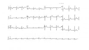
provided by Alex Voskoboinik July 2022
The Answer: Left arm – Right arm lead reversal
The emergency department doctors were concerned about T-wave inversion in lead I and aVL and diagnosed coronary ischaemia. In fact, this is a classic case of Left arm – right arm lead reversal. In this situation, Einthoven’s triangle flips 180 ̊horizontally so Lead I is inverted, aVL and aVRswitch places, as do leads II and III. The key to diagnosing lead reversals is that P waves, QRS complexes and T-waves are all inverted. In this case the p wave is negative in lead I which is not characteristic of sinus rhythm. Similarly in aVR, the p wave is positive which is not characteristic of sinus rhythm. A sinus p wave should usually be positive in all leads except aVR and is biphasic (pos/neg) in lead V1. Left arm – right arm lead reversal may appear similar to dextrocardia, however as opposed to dextrocardia there is normal precordial R wave progression in this case. This patient did not proceed to an angiogram.