Heart Failure Research Review, Issue 71, with commentary by Dr Mark Nolan

In this issue:

  • Dapagliflozin in HF with mildly reduced or preserved EF
  • Acetazolamide in acute decompensated HF with volume overload
  • Association of empagliflozin treatment with albuminuria in HF patients
  • Hyperkalaemia as a cause of MRA undertreatment in HF
  • Riociguat for pulmonary hypertension with HFPEF
  • Transvenous right greater splanchnic nerve ablation in HFPEF
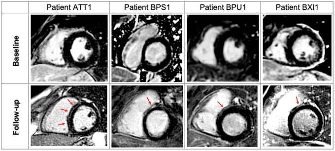
  • Stem cell-derived extracellular vesicles reduce expression of cardiac hypertrophy molecules
  • Periodontal status, CRP, NT- proBNP and incident HF
  • Alerting clinicians to 1-year mortality risk in patients hospitalised with HF
  • Uric acid and SGLT-2 inhibition with empagliflozin in HFREF

Download the Heart Failure Research Review Issue 71

Longitudinal Assessment of Structural Phenotype in Brugada Syndrome Using CMR

Longitudinal Assessment of Structural Phenotype in Brugada Syndrome Using Cardiac Magnetic Resonance Imaging

Recently published in Heart Rhythm O2 October 17, 2022  

With summary by author, Dr Julia C. Isbister 

Brugada syndrome (BrS) has traditionally been considered a channelopathy but in recent years, paralleling advancement in imaging techniques, subtle structural changes have been observed in a number BrS cohorts around the world. Cardiac magnetic resonance (CMR)imaging has revealed that patients with BrS have increased right ventricular and right ventricular outflow tract volumes compared to health controls. Focal fibrosis evidence by late gadolinium enhancement (LGE) has also been reported. Indeed, it has been postulated that BrS may be a focal cardiomyopathy rather than a pure ion channel disease.

This pilot study (n=18) represents the first time patients with BrS have been studied longitudinally with CMR and revealed that the myocardial changes can develop or progress over time. Most strikingly 22% of patients developed mid-wall LGE, typically associated with dilated cardiomyopathy, in the absence of other identifiable causes.

This work is hypothesis generating and we hope that the interesting results will prompt other groups around the world to review their patients with BrS to determine if progressive structural changes are observed in other cohorts. Ultimately, correlation of any progression of structural change with arrhythmic events and patient outcomes will be needed to determine the clinical implications of these observations and determine if serial structural assessment may aid risk stratification in BrS.

Isbister, J.C. et al, 2022, ‘Longitudinal Assessment of Structural Phenotype in Brugada Syndrome Using Cardiac Magnetic Resonance Imaging’ Heart Rhythm O2  

DOI: https://doi.org/10.1016/j.hroo.2022.10.004 

Read article in full


Abbott: Subset of Assurity™, Endurity™ and Zenex™ Pacemakers (July 2022)

A laser surface preparation may not have properly prepared the device’s metal housing potentially leading to abnormal device-to-header adhesion which may allow moisture ingress into the header. 

ANZCDACC Product Hazard Alert July 2022

  Download as pdf

Device:   A subset of Assurity™, Endurity™ and Zenex™ Pacemakers
MODELS:  PM2162, PM2172, PM2272, PM2282*
* TGA Approved but not currently commercially released
TGA Reference: 
RC-2022-RN-01000-1

ECG of the Month – July 2022

A 38 year-old develops left arm and wrist pain while riding his bicycle. ECG is shown below. A coronary angiogram is planned. What does the ECG show?

Figure 1:

provided by Alex Voskoboinik July 2022

The Answer: Left arm – Right arm lead reversal

The emergency department doctors were concerned about T-wave inversion in lead I and aVL and diagnosed coronary ischaemia. In fact, this is a classic case of Left arm – right arm lead reversal. In this situation, Einthoven’s triangle flips 180 ̊horizontally so Lead I is inverted, aVL and aVRswitch places, as do leads II and III. The key to diagnosing lead reversals is that P waves, QRS complexes and T-waves are all inverted. In this case the p wave is negative in lead I which is not characteristic of sinus rhythm. Similarly in aVR, the p wave is positive which is not characteristic of sinus rhythm. A sinus p wave should usually be positive in all leads except aVR and is biphasic (pos/neg) in lead V1. Left arm – right arm lead reversal may appear similar to dextrocardia, however as opposed to dextrocardia there is normal precordial R wave progression in this case. This patient did not proceed to an angiogram.

Coronary Artery Anomalies in Young and Middle-Aged Sudden Cardiac Death Victims

Our recent paper,  ‘Prevalence of Coronary Artery Anomalies in Young and Middle-Aged Sudden Cardiac Death Victims’ examines the rate of coronary artery anomalies in the largest population of sudden cardiac death patients examined in Australia. From a population of approximately 1500 Victorians aged 1-50 years who experienced sudden cardiac arrest, over 700 underwent a comprehensive autopsy. A 1% rate of anomalies of coronary artery anatomy was identified, which is consistent with reported rates in angiographic, CT and other post-mortem series – this is reassuring that our dataset was representative of general findings.

However, within this 1% prevalence of coronary artery anomalies, not a single person had experienced their sudden cardiac arrest due to their coronary anomaly. All patients had clear alternative reasons for their death identified, such as another coronary artery occluded with acute thrombus, histological evidence of acute myocardial infarction or a ruptured thoracic aortic dissection.

This study is important, because it challenges earlier assumptions that coronary artery anomalies are a major cause of young sudden cardiac death. Early investigations into sudden cardiac death reported that coronary artery anomalies caused up to one-third of young sudden cardiac deaths. These studies included only a few dozen patients and were published several decades ago. However, citing these studies, both US and European guidelines have traditionally restricted participation in elite sport for patients with coronary anomalies.

Our dataset is not only the largest published in Australia, but also one of the largest in the world and our findings accord with contemporary figures from other major sudden cardiac death research teams. We hope that our data will prompt a re-appraisal and further investigations into the true role of coronary artery anomalies in young sudden cardiac death.

Summary by Dr Elizabeth Paratz

Available now as a preprint in The American Journal of Cardiology
Link to full article here:


Outcomes of Thrombus Aspiration During Primary Percutaneous Coronary Intervention for ST-Elevation Myocardial Infarction

Summary by Kevin Rajakariar 15 June 2022.

Authors: Kevin Rajakariar, Nick Andrianopoulos, Daniel Gayed, Danlu Liang, Brendan Backhouse, Andrew E Ajani, Stephen J Duffy, Angela Brennan, Louise Roberts, Christopher M Reid, Ernesto Oqueli, David Clark, Melanie Freeman

The use of thrombectomy during primary percutaneous coronary intervention has been a controversial topic, with large randomised controlled trials demonstrating conflicting results. While more recent randomised studies demonstrate no clear benefit in the use of routine thrombectomy during primary PCI, there are minimal real-world studies utilising thrombectomy in selected higher risk patients. These patients may have an increased thrombus burden, higher lesion complexity, no-reflow phenomenon, and evidence of cardiogenic shock. In addition, there have been significant concerns of the peri-procedural stroke risk associated with the use of thrombectomy, with further conflicting results between studies.

Our study analysed 6,270 consecutive patients between 2007 and 2018 undergoing primary PCI for STEMI, of which 26% underwent thrombectomy. While thrombus aspiration was most likely to be used in complex lesions with no coronary perfusion, there was no significant difference in post-procedural coronary flow, stroke, or mortality. Although there was a reduction in 30-day major adverse cardiovascular and cerebrovascular events, this was not associated with a reduction in long-term mortality. However, thrombectomy was most likely to be used in sicker patients and it is unclear whether this subgroup of patients would have inferior outcomes if thrombectomy was not performed.

Available online now on the Internal Medicine Journal accepted articles page: https://onlinelibrary.wiley.com/doi/epdf/10.1111/imj.15828

Cryoablation of Papillary Muscles at Surgery for Malignant Ventricular Arrhythmias Due to Mitral Valve Prolapse

Surgical cryoablation should be considered in patients with mitral valve prolapse (MVP) undergoing mitral valve surgery who have malignant ventricular arrhythmias or high ventricular ectopy load, as recommended in our article now published online in Heart, Lung and Circulation [1].

Mitral valve prolapse is a relatively common condition with a community incidence of about 2.4% [2]. While it generally has good prognosis, a small subset of patients suffer from malignant arrhythmia and sudden cardiac death. Out of 650 autopsies in young adults who died suddenly, 7% were due to MVP [3].

Ventricular ectopics are common in MVP. In a series of 595 consecutive patients, 43% had >5% and one third had moderate or severe nonsustained ventricular tachycardia [4]. These ectopics mainly arise from one or both papillary muscles and may act as triggers for malignant ventricular arrhythmias. Radiofrequency (RF) ablation is considered for malignant arrhythmia or in MVP patients in whom high ventricular ectopy load causes LV dysfunction. RF ablation of the papillary muscles has a modest success rate and high arrhythmia recurrence rate. Mitral valve surgery by itself does not prevent malignant ventricular arrhythmia.

In Heart, Lung and Circulation, we reported 3 cases of MVP and malignant ventricular arrhythmia who not only had mitral valve surgery but also underwent cryoablation of the papillary muscles at the time of surgery.

Two patients had moderate and one severe mitral regurgitation. All had received shocks from their implanted cardioverter defibrillators (ICDs) and were not controlled with drug therapy. At surgery, encircling cryolesions were placed at the base of the papillary muscles which also targets distal arborisation of the Purkinje system. During a follow up of 3–11 years, all three patients have remained free of both malignant arrhythmia and ICD shocks. Cryoablation of papillary muscles had no detrimental effect on mitral valve function for any patient.

While we had not performed cardiac MRI in any of our patients, we would recommend it to assess areas of myocardial fibrosis which could be targeted by cryoablation. Further, wherever possible, we would now consider mitral valve repair rather than replacement.

References

  1. Vohra J, Morton JB, Morgan J, Tatoulis J. Cryoablation of papillary muscles at surgery for malignant ventricular arrhythmias due to mitral valve prolapse. Heart Lung Circ 2022. Link to online article: Cryoablation of Papillary Muscles at Surgery for Malignant Ventricular Arrhythmias Due to Mitral Valve Prolapse – Heart, Lung and Circulation (heartlungcirc.org).
  2. Levine RA, Hagége AA, Judge DP, Padala M, Dal-Bianco JP, Aikawa E, et al.; Leducq Mitral Transatlantic Network. Mitral valve disease—morphology and mechanisms. Nat Rev Cardiol 2015;12:689–710.
  3. Basso C, Perazzolo Marra M, Rizzo S, De Lazaari M, Giorgi B, Cipriani A, et al. Arrhythmic mitral valve prolapse and sudden cardiac death. Circulation 2015;132:556–66.
  4. Essayagh B, Sabbag A, Antoine C, Benfari G, Yang LT, Maalouf J et al. Presentation and outcome of arrhythmic mitral valve prolapse. J Am Coll Cardiol 2020;76:637-49.

By Jitendra Vohra, Joseph B Morton, John Morgan and James Tatoulis

ECG of the Month – June 2022

A 48 year-old male presents with 3 months of exertional dyspnoea.

He takes candesartan 8 mg daily and atenolol 50 mg daily for hypertension. ECG is shown above (Fig 1).

What is the most likely diagnosis?
Figure 1

This patient was originally diagnosed with sinus tachycardia at 100 bpm.

In fact this patient has a focal atrial tachycardia originating from the right upper pulmonary vein (ECG following ablation shown in Figure 3). Atrial flutter or atrial tachycardia with 2:1 conduction (the start of the ECG) are often misdiagnosed as sinus tachycardia. There are two important signs that should make one suspicious that this is not sinus rhythm. Firstly, the presence of a long apparent PR interval would be unusual for sinus tachycardia in a young patient whereby elevated sympathetic tone would ensure more slick conduction down the AV node. Secondly, ‘funny’ appearance of T-waves should make one suspicious that there are P waves buried within them. Looking at all leads, particularly V1 (Figure 2) and mapping them out is instructive.

Figure 2
Figure 3

Posts navigation