More Journal Articles

Pulmonary Hypertension in Aortic Regurgitation

Pulmonary hypertension (PHT) is known to complicate left heart diseases (LHD) including left sided valvular pathologies such as aortic regurgitation (AR). Patients with significant AR have an increased risk of death, even if they do not have symptoms, so identifying variables which predict adverse outcomes is important. PHT is thought to develop in AR due to left ventricular volume overload which eventually leads to increases in left atrial pressure and the development of post-capillary PHT.
Read more

Summary by Dr Seshika Ratwatte

Cardiovascular Nursing and Climate Change: a call to action from the CSANZ Cardiovascular Nursing Council

Published in Heart, Lung and Circulation, with summary by Prof Rochelle Wynne.

Read more

Exertional breathlessness is one of the commonest symptoms leading to visits to a cardiologist.

Among the myriad of causes of breathlessness, pulmonary hypertension (PH) is important and frequently under-recognised.
Our latest article on this subject was led by Dr Pyi Naing FRACP, published in the Internal Medical Journal

Read more

Prevalence and prognostic impact of tricuspid regurgitation in patients with cardiac implantable electronic devices…

Published in International Journal of Cardiology, with summary by Sophie Offen.    

Read more

The carbon footprint of hospital diagnostic imaging in Australia

Published in The Lancet, with summary by Dr Scott McAlister and Prof Alexandra Barratt.

Read more

Outcomes of Thrombus Aspiration During Primary Percutaneous Coronary Intervention for ST-Elevation Myocardial Infarction

Now available online at Internal Medicine Journal.
Summary by Kevin Rajakariar 15 June 2022.
Read more

Cryoablation of Papillary Muscles at Surgery for Malignant Ventricular Arrhythmias Due to Mitral Valve Prolapse

Published in Heart Lung and Circulation, 10 June 2022 
Summary by Jitendra Vohra, Joseph B. Morton, John Morgan, James Tatoulis.
Read more

Specialists keen for further cardio-oncology services and education

Published in Internal Medicine Journal, with summary by Prof Kazuaki Negishi, 24 April 2022
Read more


Prediction of Pacemaker Requirements in Patients with Unexplained Syncope: the DROP Score

Xiaoman Xiao et al, Heart, Lung and Circulation
Summary by co-author Alex Voskoboinik,
31 March 2022
Read more

IMPACT for Suspected ACS in ED: An Appropriate Pathway for Aboriginal and Torres Strait Islander patients too

Louise Cullen et al, published in Heart Lung and Circulation.  
Summary by Paul Bridgman, 23 March 2022.
Read more

Heart attack awareness in Australian masters football players

Published by Internal Medicine Journal 15 March 2022.
Summary by Paul Bridgman.
Read more

Statin side effects are largely nocebo

By Paul Bridgman, 22 February 2022
Read more

Pulmonary Hypertension in Mitral Regurgitation

Presented by Dr Seshika Ratwatte

Pulmonary hypertension (PHT) commonly co-exists with significant mitral regurgitation (MR), but its prevalence and prognostic importance has not been well documented. In MR, PHT is thought to be due to the direct effect of systolic backflow into the left atrium (LA), causing backpressure into the pulmonary vasculature and may develop before patients experience symptoms or left ventricular (LV) systolic dysfunction.

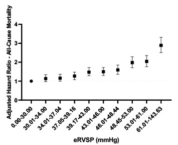
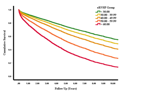
In an upcoming publication in Open Heart, Dr Seshika Ratwatte, senior author Professor David Celermajer and their co-authors identify a cohort of 9683 patients with “isolated” moderate or severe MR and preserved left ventricular ejection fraction (LVEF>50%) using the National Echo Database of Australia (NEDA). The authors show that the risk of all-cause mortality progressively increases as eRVSP level increases even after adjustment for age and sex, with aninflection’ for mortality seen from eRVSP 34mmHg onwards (Central Illustration).

The publication confirms the high prevalence of PHT in patients with significant MR and preserved LVEF on echo (59.6%). Whilst a treatment effect was not reported, it was demonstrated that even minor elevations in pulmonary pressures were associated with negative prognostic implications. Consistent with the authors work on PHT in aortic regurgitation a typical phenotype of “left heart disease” on echo was confirmed the proportion of patients with RV dilation and functional impairments as well as right and left atrial sizes increasing progressively, from no PHT to severe PHT.

Central Illustration:

Test your ECG knowledge

Figure 1

A 72 year old woman presents with palpitations and pre-syncope. Initial ECG is shown (Figure 1):

Figure 2

A Valsalva manoeuvre is attempted and subsequent ECG is shown
(Figure 2):

A 19 year-old woman presents with palpitations. Continuous rhythm strip post adenosine is shown. What is the likely mechanism of tachycardia? Does this patient need an urgent ablation?

A/Prof Alex Voskoboinik, March 2023

Read more and find your answer

A 68 year old male with a long standing non-ischaemic cardiomyopathy is admitted with a NSTEMI and found to have triple vessel disease. On Day 1 post-CABGs, he complains of dizzy spells and the following is seen on telemetry.

A/Prof Alex Voskoboinik, February 2023

Read more and find your answer

A 16 year old girl with a history of recurrent paroxysmal palpitations and a structurally normal heart has the following 12-lead ECG recorded during an especially severe episode for which she obtained urgent medical assistance. 

Haris Haqqani, November 2022 

Read more for diagnoses and explanation:


A 57 year-old woman with a loop recorder in-situ for investigation of syncope presents to the emergency department with recurrent dizzy spells. Loop recorder tracings (1 & 2) are shown on the link below

Read more and find your answer

Alex Voskoboinik  October 2022

A 76 year-old gentleman presents for a pacemaker check 4 months post implant. High ventricular rate episodes are noted, as shown below. He is asymptomatic during episodes:

Read more and rest your answer 

Alex Voskoboinik  September 2022

A 28 year old nurse with a long history of paroxysmal palpitations has the following 12-lead ECG and 2-channel telemetry trace recorded during an episode.

Read more and find your answer

Haris M. Haqqani August 2022

A 38 year-old develops left arm and wrist pain while riding his bicycle.  A coronary angiogram is planned. What does the ECG left show?

Click here to find the answer 

Alex Voskoboinik July 2022

A 48 year-old male presents with 3 months of exertional dyspnoea.
He takes candesartan 8 mg daily and atenolol 50 mg daily for hypertension. ECG is shown left:

What is the most likely diagnosis?        Read more here to find the answer…

Alex Voskoboinik June 2022

Stem: A 29 year-old male presents with a 2 month history of frequent palpitations. Bedside TTE shows mild-moderate global LV dysfunction. What are the differential diagnoses?

Click here to read more and to find the answer 

Alex Voskoboinik May 2022

A 25 year-old male with no past history presents with palpitations.

What is the most likely diagnosis?    Why are there two QRS morphologies?

Alex Voskoboinik April 2022

Click here to read more and find the answer

ECG of the Month, presented by A/Prof Alex Voskoboinik

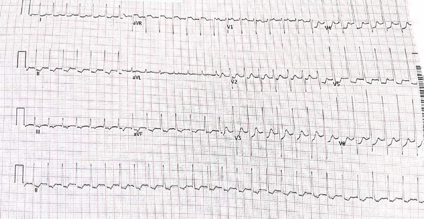
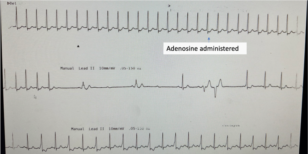
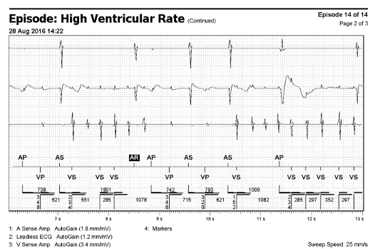
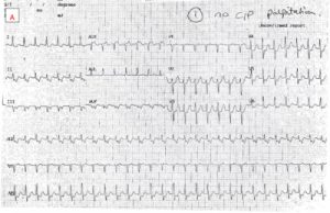
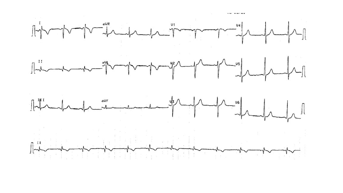
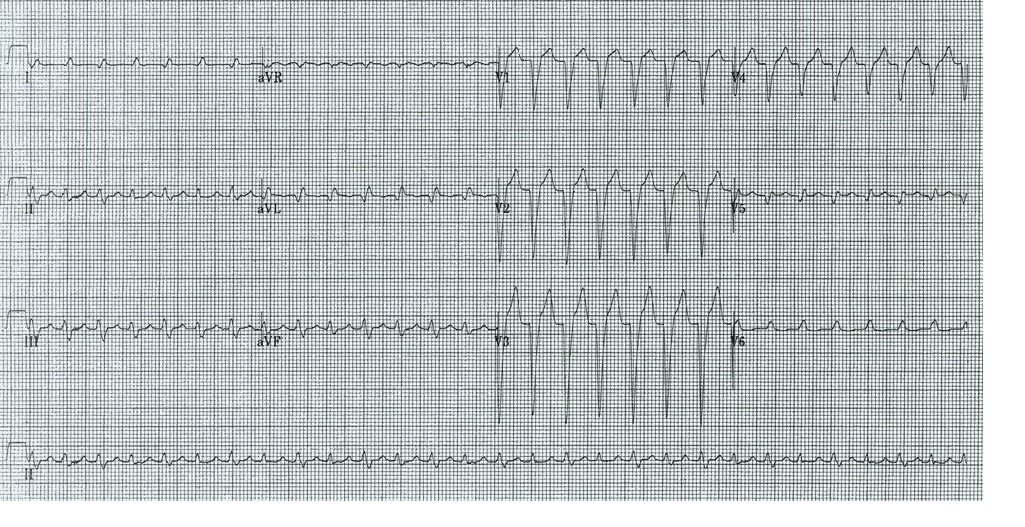
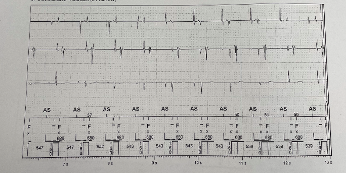
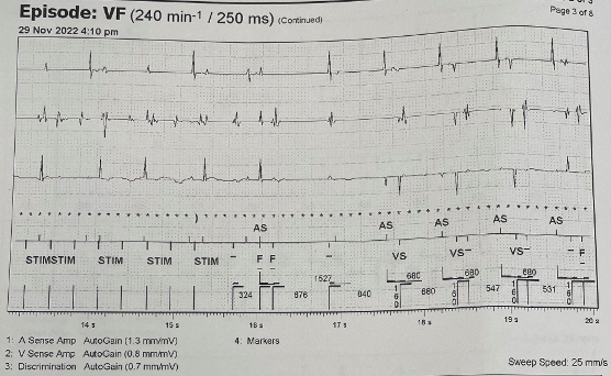
A 72 year old woman presents with palpitations and pre-syncope. Initial ECGs is shown below (Figure 1):

Figure 1

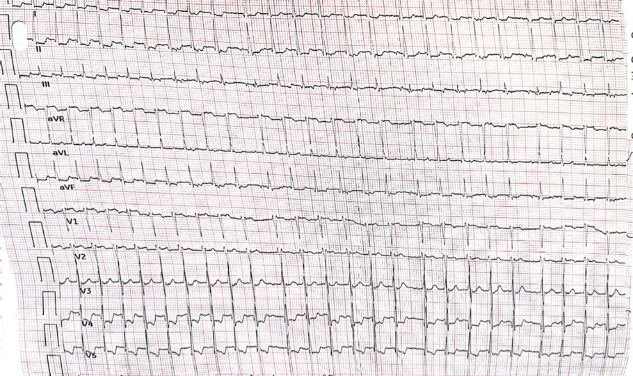
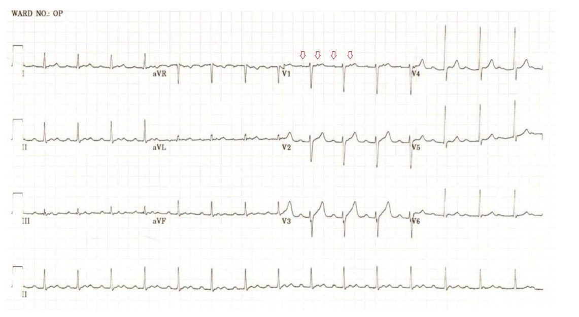
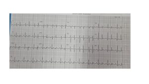
A Valsalva manoeuvre is attempted and subsequent ECG is shown below (Figure 2):

Figure 2

What is the most likely diagnosis? Want to ask a question or discuss visit the ECG Forum…


Answer:

Atrial tachycardia with Wenckebach AV nodal conduction.

Explanation:

The initial ECG demonstrates a ‘short RP’ SVT with the p-wave best seen in lead aVL on the initial ECG. The differentials are  typical atrioventricular nodal reentrant tachycardia (usually the retrograde p wave is buried at the end of the QRS which is not the case here), atrioventricular reentrant tachycardia or atrial tachycardia (AT) with first degree AV block.

The second ECG following a ‘vagal’ manoeuvre does not terminate the tachycardia but increases the block at the level of the AV node. This exposes the p-waves and there are more ‘A’s than ‘V’s with grouped beating seen (best appreciated in lead III). This Wenckebach pattern excludes AVRT (in fact by definition the A:V relationship must be 1:1) and makes AVNRT very unlikely. The p-wave morphology is nearly isoelectric in V1 and very ‘skinny’ suggesting a mid-line origin. In pact at EP study the focal atrial tachycardia was mapped to the perinodal region and successfully ablated there.

Extensive pulmonary artery embolisation and pulmonary hypertension caused by cardiac hydatid cyst rupture

Extensive pulmonary artery embolisation and pulmonary hypertension caused by cardiac hydatid cyst rupture

Dr Jason Harmer

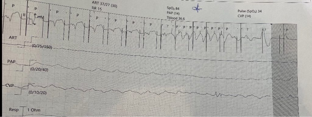
This case is a rare example of cardiac hydatidosis found in a patient in rural NSW, Australia. He was a middle-aged man who initially presented with a ruptured right ventricular hydatid cyst causing an anaphylactoid-like reaction, pulmonary emboli and widespread dissemination of hydatid echinococcus throughout the lung. He survived the cyst rupture and underwent cardiac surgery, but had incomplete resection and experienced progressive cardiopulmonary hydatidosis despite ongoing antihelminthic therapy. He had an array of cardiopulmonary sequelae over his lifespan, including worsening pulmonary hypertension and right heart failure, as well as bone marrow suppression and neutropenic sepsis. This case highlights rare clinical manifestations of cardiac hydatidosis and potential complications of its treatment.

Hydatidosis is caused by ingestion of larvae from the Echinococcus granulosus tapeworm from contaminated food or water supply or by direct contact with infected animals. Hydatid cysts commonly affect the liver or lung; however, cardiac involvement can occur in rare cases (i.e. <2% of all cases of hydatidosis).

Figure 1
Transthoracic echocardiogram: apical 4 chamber view. Intracardiac hydatid cyst.
Apical attachment of a large hydatid cyst to the wall of the RV. The RV chamber is dilated and hypokinetic and the septum is dyskinetic. The distal portion of the hydatid cyst is mobile. The RA is severely dilated and the interatrial septum is displaced due to pressure overload. The tricuspid annulus appears dilated.

Figure 2
Transthoracic echocardiogram: parasternal short axis view. Intracardiac hydatid cyst.

There is a large mass (hydatid cyst) in the RV that appears to be fixed to parts of the interventricular septum. Motion of the interventricular septum is abnormal, in part due to attachment of the mass and from raised pressures in the right heart. Interventricular septal flattening is a sign of raised right heart pressures. This patient has known moderate to severe pulmonary hypertension as a consequence of dissemination of hydatid disease to the lung and pulmonary thromboembolism.
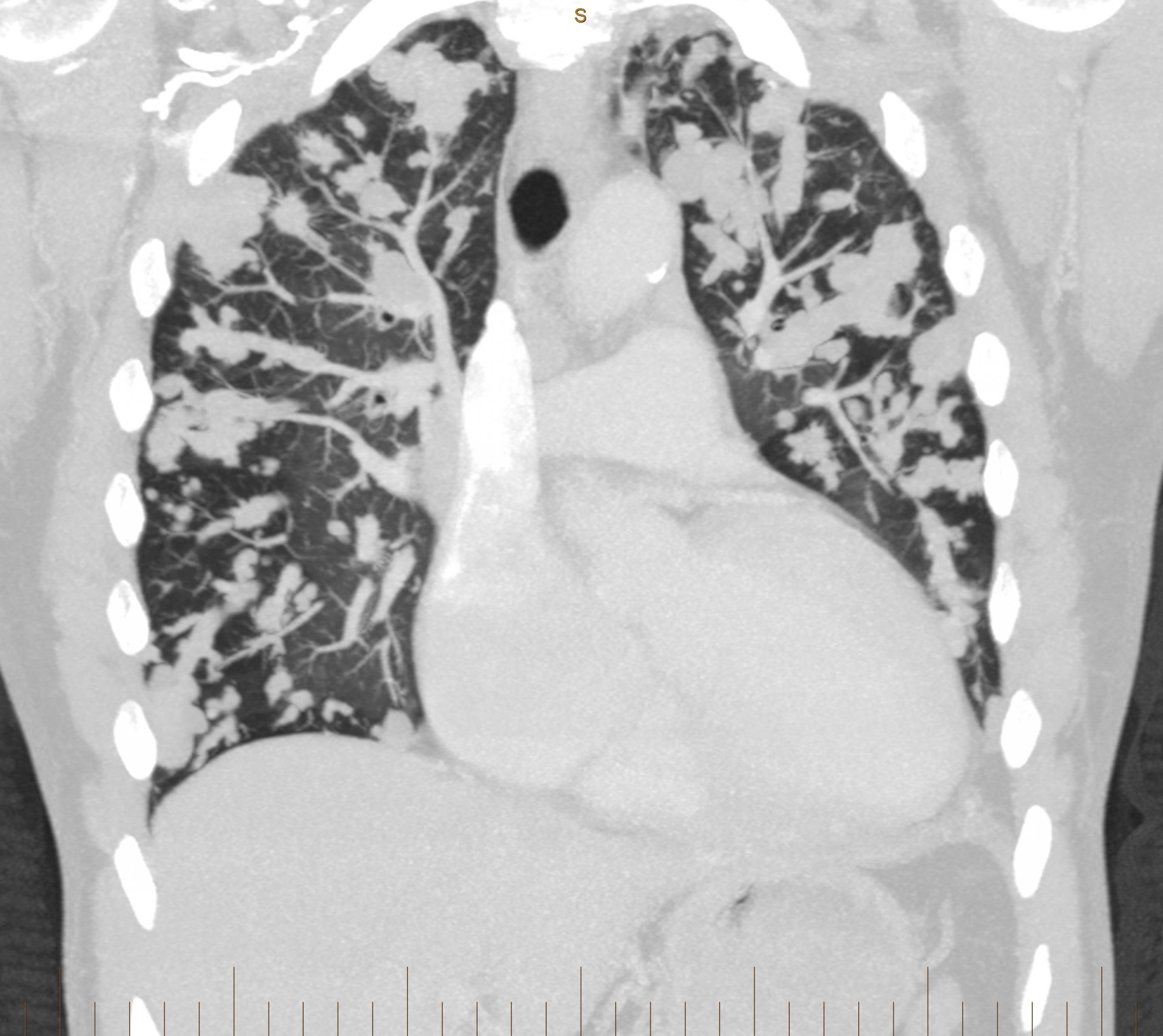
Figure 3

Figure 3
CT pulmonary angiogram: coronal view. Multiple hydatid masses within the pulmonary vasculature.
Multiple lobulated areas within the pulmonary vasculature from extensive dissemination of hydatid disease originating from the RV.
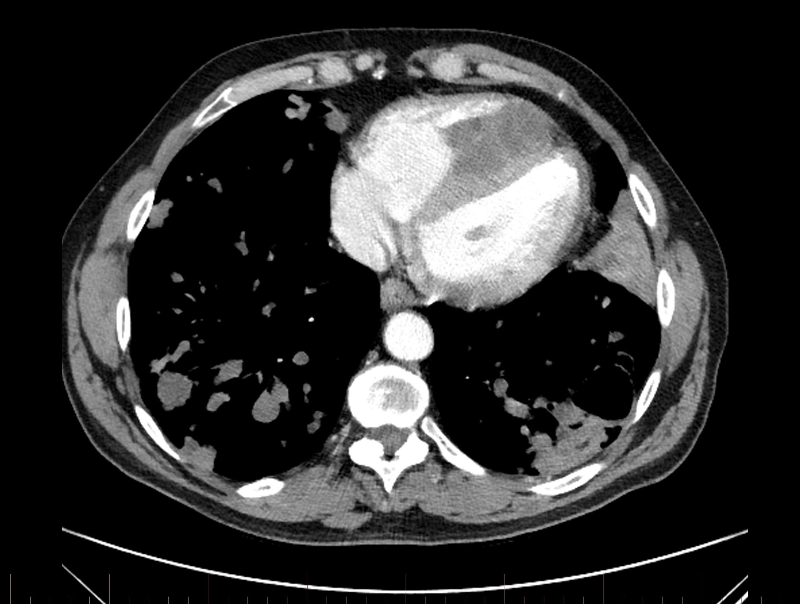
Figure 4

Figure 4
CT pulmonary angiogram: axial view.
There is a dense mass within the RV chamber due to a hydatid cyst. There is reduced RV blood filling in diastole due to the space occupying mass.  The interventricular septal wall is clearly demarcated and the mass appears to encroach on the septal wall at two points. The RV is enlarged and occupies the apex of the heart. The sternum has been divided in two parts from a previous stenotomy (with incomplete surgical resection of the mass, leading to regrowth). There is a cavitating lesion in the posterior aspect of the left lung.

Abbrev. CT = computed tomography; RV = right ventricle; LV = left ventricle; RA = right atrium.


Pulmonary Hypertension in Aortic Regurgitation

Pulmonary Hypertension in Aortic Regurgitation
by Dr Seshika Ratwatte

Pulmonary hypertension (PHT) is known to complicate left heart diseases (LHD) including left sided valvular pathologies such as aortic regurgitation (AR). Patients with significant AR have an increased risk of death, even if they do not have symptoms, so identifying variables which predict adverse outcomes is important. PHT is thought to develop in AR due to left ventricular volume overload which eventually leads to increases in left atrial pressure and the development of post-capillary PHT.

Now published in BMJ Heart, Dr Seshika Ratwatte, senior author Professor David Celermajer and their co-authors identify a cohort of 8392 patients with “isolated” moderate or severe AR and preserved left ventricular ejection fraction (LVEF>50%) using the National Echo Database of Australia (NEDA). The authors show that the risk of all-cause mortality increases as eRVSP level increases even after adjustment for age and sex (Central Illustration), with a ‘threshold’ for mortality seen from mild PHT onwards.


The publication outlines that a significant proportion of the population with significant AR had suspected PHT based on echo. Echo remains the first line screening tool to detect PHT and is the guideline recommended diagnostic method of choice to allow for monitoring and follow up. Clinicians need clear parameters to monitor and prognosticate such patients accurately. The authors identified a typical phenotype of “left heart disease” on echo with E:e’, right and left atrial sizes and the proportion of patients with RV functional impairments increasing progressively, from no PHT to severe PHT.

Central Illustration:


Link to publication is now available in BMJ Heart. https://heart.bmj.com/content/early/2023/04/02/heartjnl-2022-322187

ECG of the Month Feb 2023

ECG of the Month
presented by A/Prof Alex Voskoboinik

A 68 year old male with a long standing non-ischaemic cardiomyopathy is admitted with a NSTEMI and found to have triple vessel disease. On Day 1 post-CABGs, he complains of dizzy spells and the following is seen on telemetry:

The most likely explanation is:

  1. Artefact
  2. Ventricular tachycardia
  3. SVT with aberrancy
  4. Oversensing
  5. Undersensing

The most likely explanation is:

  1. Artefact
  2. Ventricular tachycardia
  3. SVT with aberrancy
  4. Oversensing
  5. Undersensing
Explanation

This patient has a pre-existing ICD (which remains active) and epicardial pacing wires. The beginning of the telemetry strip starts with A-V pacing from the epicardial pacing leads with a large pacing stimulus visible from stimulation of both chambers. The ICD then proceeds to deliver anti-tachycardia pacing (which appears as the wide complex ‘tachycardia’ with pacing spikes prior to each QRS complex). The haemodynamic effects of this ATP can be seen on the PAP (Pulmonary Artery Pressure) invasive waveform with flattening of the trace seen. The ICD interrogation is shown, and demonstrates oversensing of the epicardial pacing stimulus on the ICD’s ventricular channel – hence the ICD double counts both the stimulus and the ventricular paced beat and hence concludes that the patient is in VT/VF, and delivers anti-tachycardia pacing.

Discuss or post a question on the CSANZ Forum 

Cardiovascular Nursing and Climate Change: a call to action from the CSANZ Cardiovascular Nursing Council

Cardiovascular Nursing and Climate Change:  a call to action from the CSANZ Cardiovascular Nursing Council.

Summary by Prof Rochelle Wynne December 2022

Environmental health ultimately determines human health. As our climate continues to deteriorate, climate hazards and disasters provide acute triggers for patients with cardiovascular disease (CVD) that are compounded by social determinants of health.

Nurses provide a critical juncture for future solution focused co-design. To avoid harm, we need to have a holistic view of vulnerability, recognise what influences resilience, and appreciate the synergistic effects of climate change on CVD.

In a special issue of Heart, Lung and Circulation dedicated to the environment, this call-to-action claims that unprecedented should not be an excuse for unprepared. In harnessing the power, skill and expertise of the nursing profession, healthcare professionals who are both part of the solution and the problem, can transect acute and primary care to lead positive change.

Vulnerable populations are at greatest risk and targeted preventative support is needed. First Nations people must have sovereignty over their response to climate change. Healthcare professionals need to understand the impact of climate change on First Nations peoples and ensure care is culturally appropriate.

Education is the cornerstone to mitigating the impact of climate change on healthcare outcomes as it creates opportunities to reduce the risk of hazards and establish strategies for safety.

Urgent practice adaptation that includes climate-aware assessment, education, discharge planning and care coordination as usual practice is imperative. Professionals who manage the consequences of climate change must also understand the impact of their care on the root cause of the problem. As we adapt our care to reduce environmental impact nurses are well placed to lead interdisciplinary planning and this paper highlights priorities for action to improve awareness and empower self-care in climate related disasters.

Read in full at Heart, Lung and Circulation here – https://www.heartlungcirc.org/article/S1443-9506(22)01137-4/fulltext 

Authors: Sally C. Inglis, PhD, Caleb Ferguson, PhD, Rebecca Eddington, MHLth, Julee McDonagh, PhD, Chris J. Aldridge, Kimberley Bardsley,  Dion Candelaria, MN, Y.Y. Chen, PhD,  Robyn A. Clark, PhD, Elizabeth Halcomb, PhD, Jeroen M. Hendriks, PhD.  Louise D. Hickman, PhD, Rochelle Wynne, PhD.

Posts navigation